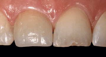
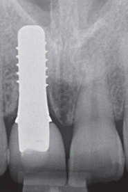
Mobirise

CLINICAL CASES - B

ZERAMEX® XT - Immediate Placement: site # 21

Courtesy Prof. Dr. med. Dr. med. dent. Michael Stiller & Dr. Barbara Peleska
| [email protected] | www.implant-consult.de | [email protected]
( click on the title above to access the published article in a new window)
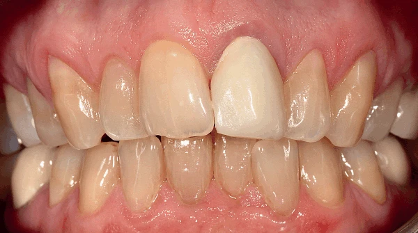
1- : Clinical situation: Narrow jaw with an overjet of 9 mm. Buccal view.
2- : Clinical situation: Narrow jaw with an overjet of 9 mm. Lateral view.
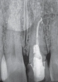
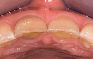
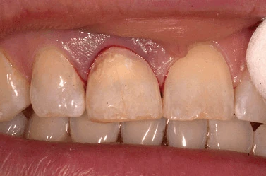
3- Tooth 21 had a massive palatal and sub-
gingival carious lesion
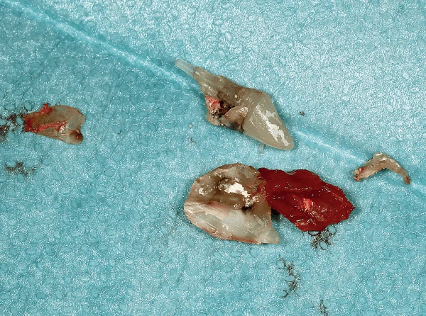
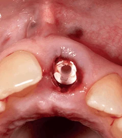
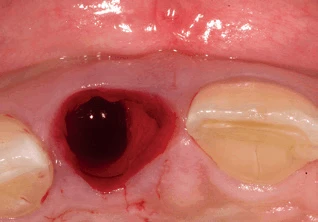
4- Tooth 21 removed.
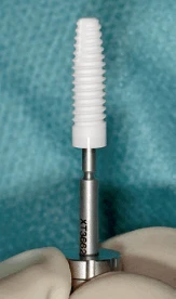
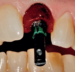
5-Zeramex XT RB 4,2mm x 14mm implant - mounted on a Zeramex pickup, ready for insertion
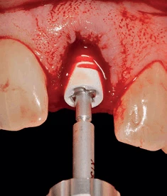
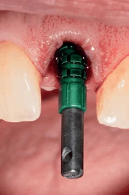
6- XT implant inserted into prepared alveola with Zeramex pickup instrument.
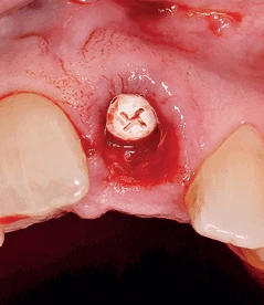
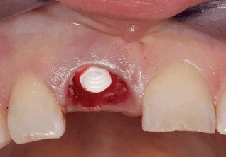
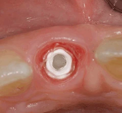
7-Implant in situ with good primary stability.
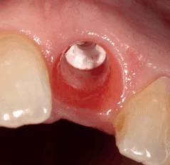
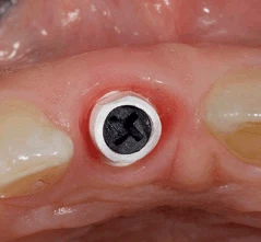
8- Healing cover
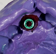
9- Difference between buccal and palatal gingival margins due to overjet.
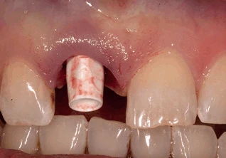
10- Transfer sleeve inserted to check axis.
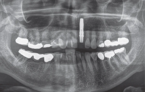
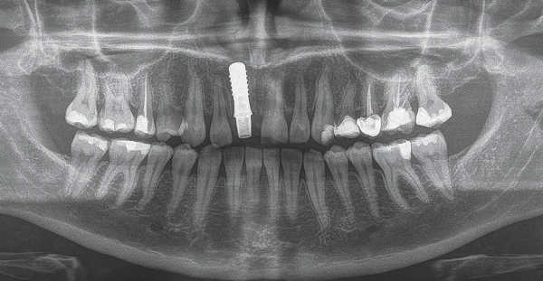
11- Control OPG - post-op | XT with RB straight abutment.
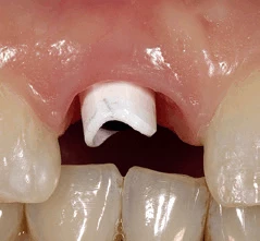
12-Temporary crown bonded to RB straight abutment.
13-Temporary crown screwed in place with Vicarbo screw.
14-Site # 21 after 6 months- ready for final impression.
15- Modified open transfer sleeve for final impression.
16- VPS impression with transfer sleeve ready to be sent to laboratory.
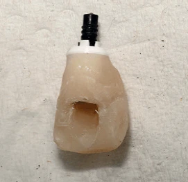
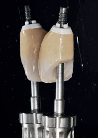
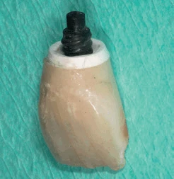
17- Final crown/abutment assembly with Vicarbo screw mounted on prosthetic key.
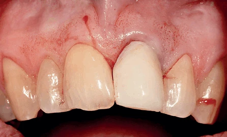
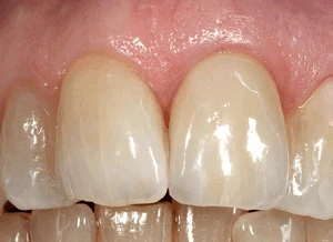
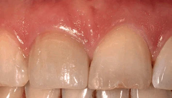
18- Final restoration # 21 - buccal view. Note excellent soft tissue appearance and colour.
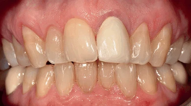
19- BEFORE
20- AFTER - Buccal view:  Fully reversible final restoration on # 21, screwed in place without any cement.
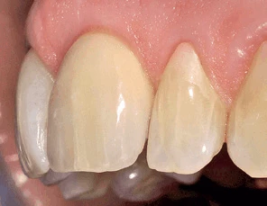
21- AFTER - Lateral view:  Excellent emergence profile, soft tissue contour and excellent condition, especially compared to the initial situation.

 ZERAMEX® P6
 implants in the aesthetic anterior region 
- Immediate placement: Site # 11

Courtesy Prof. Dr. med. Dr. med. dent. Michael Stiller & Dr. Barbara Peleska
| [email protected] | www.implant-consult.de | [email protected]
( click on the title above to access the published article in a new window)
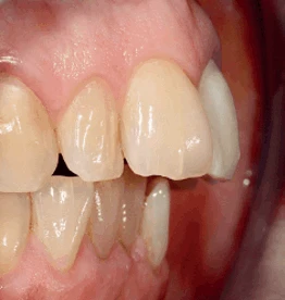
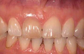
1- Initial situation
2- initial situation.
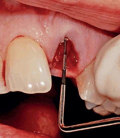
3-The fractured tooth could be removed atraumatically without bone loss after removal of the provisional crown.      
4-After the osteotomy the implant was positioned slightly palatally with primary stability.
5-Screwed zirconia abutment.
6- OPG control image.
7-The temporary crown was made with a light-curing composite bonded to the straight zirconia abutment
8- Temporary crown in place post-operatively.
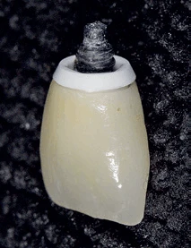
9- Final crown/abutment complex.
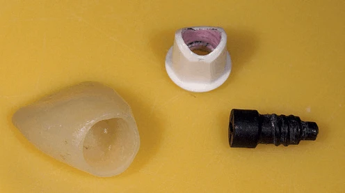
10- Crown, abutment and Vicarbo screw.
11- P6 implant prior to screwing the abutment.
12- Abutment in place.
13- buccal view of the abutment.
14- Crown # 11 in place
15- The clinical picture  two years post-op show peri-implant conditions without inflammation and excellent soft tissue health.
16-  X-ray inspection two years post-op show peri-implant conditions without inflammation